Kinesiology Taping, Part Two
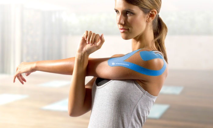
Are you using kinesiology tape properly to treat pain and reduce disability? Registered osteopath John Gibbons of Athletics Weekly provides some general guidelines on how to use kinesiology tape as well as specific techniques for taping two common injuries: shin splints and patellofemoral pain syndrome.
General rules
Before application:
- Always check for a history of allergies to tape adhesives
- Cleanse skin from any oil, cream and massage wax, and trim hair if needed
- Measure and cut the tape into the size and shape required
- Round off the corners at the end of each tape to prevent it from lifting or peeling
- Never stretch the ends of the tape and always leave around 2-3cm of tape at each end that will remain unstretched. Leaving no stretch at the ends of the kinesiology tape will avoid a “shearing” type of tension to the skin and will limit any potential for irritation as the tape is normally kept on for at least a few days.
Pre-stretch and tape application:
Before the kinesiology tape is applied to the area that is injured, guide and place the soft tissue of your patient, such as the muscle, into a position that will cause the tissue to be naturally stretched. Prior to applying the kinesiology tape, expose the adhesive side of the tape so that it can be attached to the specific body area. It is natural to want to peel off the backing from the tape—however, this process is not needed as the tape can simply be torn across one of the squares on the back. This tearing will not damage the kinesiology tape, as only the backing will be removed.
Apply a prepared ‘I’ or ‘Y’ strip to the pre-stretched tissue of the body, with little to no stretch of the tape on first application. This technique will help stabilize the area.
Treating shin splints
Athletes can often have pain that can be localized to the lower medial aspect of the tibia, especially straight after or during sporting activity. The condition starts as an irritation of the outer lining of the bone called the periosteum and can lead to periostitis. The muscles that are normally responsible for this type of pain at the medial aspect of the tibia are the tibialis posterior, flexor digitorum longus and flexor hallucis longus.
If left untreated, the medial tibia can stress the bone and eventually lead to a stress fracture. In the worst case, if this injury is neglected, a posterior compartment syndrome can develop and a surgical fasciotomy to reduce the pressure within the myofascial compartment might be recommended.
The patient adopts a long sitting position and is instructed to dorsiflex their ankle and evert their foot to place the tibialis posterior on stretch.
Apply an ‘I’ strip from just below the medial malleolus and ideally attach the tape from the navicular bone.
Apply the tape with little to no stretch.
Follow the medial shin so that the area of pain is covered.
Apply a ‘Y’ strip and start posterior to the pain with a 75% stretch to each tail of the tape.
Apply across the hotspot of the painful area. You can activate the stickiness of the glue, by rubbing and warming the tape briskly.
Treating patellofemoral pain syndrome (PFPS)
Patellofemoral pain syndrome (PFPS) is a painful condition that can relate to a type of mal-tracking of the kneecap (when the muscles pull the patella tendon in different directions).
This condition has many causes, such as an overpronation of the subtalar joint (STJ) of the ankle and poor foot biomechanics.
Weakness of the inner quadriceps muscle can also contribute to PFPS, especially the vastus medialis oblique (VMO) fibers, which are thought to atrophy due to pain and minimal swelling. In addition, weak gluteus medius and gluteus maximus can cause this type of knee pain.
The knee joint is therefore what I refer to as “a weak link in the kinetic chain” and typically the presentation of the pain is not where the problem lies.
The patient is asked to adopt a long sitting position with their knee at 90° of flexion.
Next attach a ‘Y’ strip from the superior aspect of the patella and apply the tape, with no stretch, to the medial and lateral sides of the patella. Finish by crossing over the tibial tuberosity.
Apply a ‘Y’ strip from the tibial tuberosity and lay the tape medially and laterally around the patella so that it overlaps the first ‘Y’ application. Apply with little to no stretch and finish near the quadriceps tendon.
Heat to activate the glue and once the glue has been heat-activated lower the limb back down to the couch and observe the “wrinkling” of the tape. This illustrates the effect the kinesiology tape is having on the underlying soft tissues through its unique lifting motion.


.webp)

