Soft Tissue Treatment in Athletic Injuries
Over the past decade, athletic trainers have begun to significantly expand their practices to include certain forms of injury treatment. Even professional athletes have begun to benefit from soft tissue manipulation by sports chiropractors and athletic trainers. In an article in Dynamic Chiropractic, Joseph M. Horrigan, DC, DACBSP, examines how chiropractic knowledge of the process of soft tissue injuries can help athletic trainers better work with athletes and provide helpful early interventions.
Injury & Healing Process
A significant level of interest and investigation into the injury and healing  processes of soft tissue structures has taken place over the last 10 to 15 years. This has shed light on the nature of the end result of the injury, which may include limitations in range of motion, decreased strength and overall function that follows a soft tissue injury.
The field of chiropractic and athletic training has had its origin and main focus in joint manipulation. The evolution of the clinical science database has given us greater understanding of the soft tissue injury. As practicing doctors that are treating athletes, this information allows us to accomplish two critical points:
- Understand the torque that soft tissue fibrosis and adaptive shortening can exert on a joint (i.e., vertebral hypomobility and/or "subluxation"). This may be a significant factor in the failure of manipulation of vertebral segments to maintain or "hold."
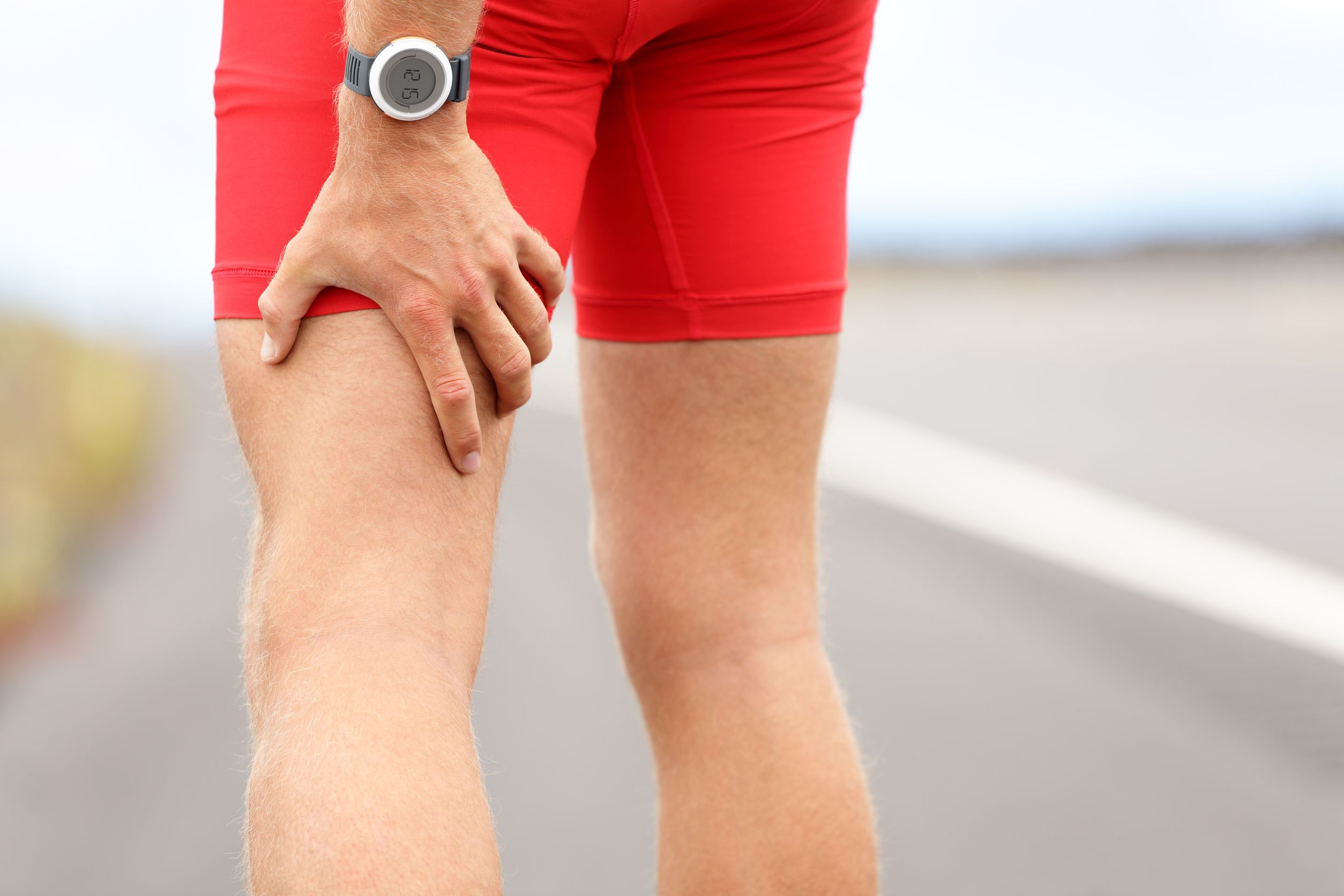
- Treatment of primary soft tissue injuries that may be unrelated to traditional chiropractic theory. This may include hamstring injuries, rotator cuff injuries, etc. The sports chiropractor is taking a more prominent role in both the treatment of athletic injuries at all levels and also in the increasing opportunity to have access to elite athletes. We have the ability to add the appropriate treatment of soft tissue injuries to our clinical acumen.
 Stages of Healing
The process of injury-healing is well documented.1-6,8-9 Examination of the injury on a histochemical level reveals multiple stages of healing, that a simple overview demonstrates:
- The injured muscle breaks up into fragments and the cellular contents are disrupted
- Satellite cells survive the degeneration and become activated
- Macrophages invade the area and remove all trace of damaged muscle and may stimulate neovascularization
- Other components of early inflammatory stages include hematoma, histamines, bradykinins, prostaglandins, hyaluronic acid, PMNs
- Fibroblasts respond to the macrophages and follow the fibrin network that was created in the wound. Myofibroblasts begin to produce wound contraction four days after the injury
- Filaments are laid down in disorganized manner (more on this later)
- Fibroblasts synthesize GAG which, together with water, form a ground substance that provides a medium for moving collagen fibers
- Early cross-links of the scar are ionic bonds and are weak. Later the bonds become covalent bonds and are much stronger
- Regenerating muscle is similar in structure to normal skeletal muscle fibers and it is responsive to its mechanical environment (more on this later)
- Remodeling of the scar tissue can last for one year.
We can see that the wound contraction begins only four days after the injury. The fibroblasts begin laying down fibrous tissue at that time and collagen fibers follow later. The scar tissue becomes much stronger in time and the remodeling can go on for one year. This is a far cry from the six-week time frame that was believed by health care practitioners for so many decades. 
Other parameters surrounding injury became evident with further research. Immobilization causes a loss of the ground substance, and that causes more cross link formation. This makes the scar tissue less mobile. The amount of scar tissue that has to be remodeled is inversely proportional to the return to function. The remodeling is responsible for the final orientation and arrangement of collagen fibers. It is the physical weave of the adhesions that will determine its mobility or restriction.
Exercise has been found to enhance the recovery ability of functional capabilities of regenerating muscle. Other remodeling factors include: muscle tension; soft tissue loading and unloading; splinting; temperature changes; and mobilization. An investigation5 was performed on rats that had muscle injuries imposed upon them. Some rats were immobilized and a second group was allowed to move (limp) in their cages according to their own ability to move.
Microscopic examination of the injury sites revealed that the rats that were allowed to move about freely had collagen laid down between the wound gap only 14 days after the injury. It is also significant to note that the collagen fibers were parallel to the muscle fibers. The connective tissue formation across the injury gap allowed the injured limb to be used before the healing was complete.
What does this all mean and how can it apply to a practice? An investigation7 found that facilities that dealt with soft tissue sports injuries and provided early intervention, rapid mobilization and aggressive exercise programs had superior results when compared to prolonged bed rest, inactivity, lengthy waiting periods before beginning physical therapy, and too many passive modalities. 
As we enter the clinical sports medicine arena with orthopedic surgeons, physiatrists, physical therapists and athletic trainers, we all must have an understanding of the injury itself. And as we enter this arena, we assume the significant medical-legal consequences of managing cases of elite athletes with multi-million dollar contracts. The understanding of the injury, keeping up with current literature of soft tissue injuries and management, acquiring contemporary skills of soft tissue injury management and diagnosis, are the tools that will not only give us to opportunity to enter this sports medicine arena, but will also keep us in the arena.
This article, originally published in Dynamic Chiropractic, was written by Joseph M. Horrigan, DC, DACBSP and originally appeared here.
References
- Carlson BM, Faulkner JA. The regeneration of skeletal muscle fibers following injury: A review. Medicine and Science in Sports and Exercise 1983, 15 (3):187-198.
- Fleckenstein JL, Shellock FG. Exertional muscle injuries: Magnetic resonance imaging evaluation. Topics in Magnetic Resonance Imaging 1991, 3:50-70.
- Fleckenstein JL, Weatherall PT, Parkey RW, Payne JA, Peshock RM. Sports-related muscle injuries: Evaluation with MR imaging. Radiology 1988, 172:793-798.
- Hardy MA. The biology of scar formation. Physical Therapy 1989, 69: 1014-1024.
- Hurme T, Kalimo H, Lehto M, Jarvinen M. Healing of skeletal muscle injury: an ultrastructural and immunohistochemical study. Medicine and Science in Sports and Exercise 1991, 23 (7):801-810.
- Lehto M, Jarvinen M, Nelimarkka O. Scar formation after skeletal muscle injury. Archives of Orthopaedic and Traumatic Surgery 1986, 104:366-370.
- Mitchell RI, Carmen GM. Results of a multicenter trial using an intensive active exercise program for the treatment of acute soft tissue and back injuries. Spine 1990, 15 (6):514-521.
- Russel B, Dix DJ, Haller DL, Jacobs-El J. Repair of injured skeletal muscle: a molecular approach. Medicine and Science in Sports and Exercise 1992, 24 (2):189-196.
- Woo AL, Buckwalter JA. Injury and repair of the musculoskeletal soft tissues. Journal of Orthopaedic Research 1988, 6:907-931.


.webp)


